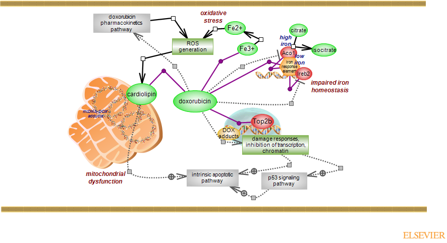
Doxorubucin (DOX, trade name Adriamycin) is an anthracycline derived from Streptomyces bacterium Streptomyces peucetius var. caesius. DOX is used in the treatment of several solid cancers, leukemia and lymphomas. The drug is composed of a planar aromatic ring with an anthraquinone chromophore and a sugar group. Once taken up by the cell and inside the nucleus for which it has a high affinity, the planar ring structure intercalates between adjacent DNA base pairs, preferentia
lly GC base pairs. DOX can form DNA adducts, binds to DNA and topoisomerases and inhibits DNA replication and transcription. It promotes DNA double-strand breaks (DSBs) and the DNA damage prompts various responses, including cell cycle arrest and apoptosis, primarily via activation of p53 pathway but also independent of it, with beneficial, if tumor cells, or otherwise detrimental consequences. While toxic to the tumor cell, DOX is also toxic to all other cell types. The cardiotoxicity of the drug is the main and the most severe adverse effect, and the cardiomyopathies associated with DOX can eventually lead to heart failure. The formation of reactive oxygen species (ROS), impaired iron homeostasis and mitochondrial dysfunction are thought to underlie the cardiotoxicity of DOX. Although more than 50% of DOX is eliminated unchanged, the drug undergoes several types of transformation such as two- and one-electron reduction and to a smaller extent deglycosidation. Free radical generation is a feature of DOX metabolism. The drug can interact with iron and generate hydroxyl radical and with the iron-responsive elements (IRE) regions of mRNAs, it can impact on the function of iron regulatory proteins (IRPs) and disturb iron homeostasis. In addition, DOX localizes to mitochondria, forms adducts with mitochondrial DNA, interacts with cardiolipin - an essential component of inner mitochondrial membrane, and is capable of impacting on a number of proteins/enzymes important for mitochondrial function. Doxorubicin is also administered as a pegylated liposomal product.
DOX has a high affinity for iron and forms 1:1, 2:1 and 3:1 drug-metal complexes. DOX binds the ferric iron (III) which can then be reduced to ferrous iron (II) to further give rise to the toxic hydroxyl radical species that can induce lipid peroxidation, nucleic acid and protein damage. As mentioned, reactive oxygen species (ROS) are also a by-product of DOX metabolism. DOX binds IREs (iron response elements), hairpin structures at the 5' and/or 3' untranslated mRNAs and found in various proteins important for iron homeostasis. The IREs are recognition sites for the iron regulatory proteins (IRPs): Aco1 and Ireb2. Binding to the 5' IRE of ferritin - the iron storage protein, inhibits its translation. In the case of transferrin receptor (Tfr1) - the major iron uptake protein, IRE is 3' located and binding of IRPs increases its translation. Aco1 is a dual function protein which can act as an aconitase to convert citrate to isocitrate if an iron-sulfur cluster [4Fe-4S] is present, as is the case when intracellular iron levels are high (holo-IRP1/Aco1). In iron-depleted cells the cluster is absent (apo-IRP1/Aco1) and Aco1 is now an RNA binding protein, binding the IREs. Ireb2, known as IRP2, does not contain an iron-sulfur cluster. DOX impairs the function of the two regulators but the actual molecular mechanisms are still a matter of debate.
Cardyomyocytes, due to the high energy demand, have a large number of mitochondria and as such, mitochondrial dysfunction can elicit large and negative effects. Like with the nuclear DNA, DOX can form adducts with mitochondrial DNA. In addition, DOX binds cardiolipin, the important lipid of the inner mitochondrial membrane(IMM) where it accounts for ~25% of its content. It is commonly referred to as the signature phospholipid of mitochondria. Cardiolipin interacts with a large number of proteins including components of the electron transport chain complexes, enzymes and metabolite carriers. The synthesis of cardiolipin is a complex metabolic system involving both a de novo arm and several remodeling routes and it is strictly local, taking place in the mitochondria. While the remodeling of cardiolipin may involve its translocations, the presence of cardiolipin at the outer mitochondrial membrane (OMM) can constitute a damage signal leading to mitochondrial autophagy (mitophagy) and apoptosis. Mitophagy can exert an early cardioprotective response by removing dysfunctional mitochondria. Increased stress however will lead to cell death.
Cardiomyocytes appear to preferentially express Top2b, and the 'trapping' of the enzyme within the DNA cleavage complex which blocks the resealing of strands, leads to various damage and other responses culminating in cell death, desirable if the cell is a tumor cell, but here detrimental. ROS are normal byproducts of mitochondrial respiration and given the large presence of mitochondria, their production/presence is likely higher than in other cell/tissue types. Interestingly however, cardiomyocytes exhibit lower antioxidant responses and have lower levels of antioxidant enzymes. For these and all other reasons presented above, the specific and extremely cardiotoxic effect of doxorubicin may not necessarily be surprising. Iron chelators and cardiomyocyte-specific Top2b deletion in a mouse model, have shown protective effects against doxorubicin-induced cardiotoxicity. To see the ontology report for annotations, GViewer and download, click here