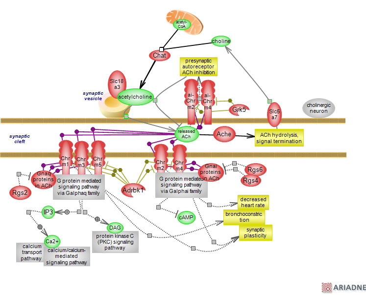
Acetylcholine (ACh) is a major neurotransmitter and neuromodulator that plays important roles in the central and peripheral nervous systems, the autonomic nervous system (ANS), and also in non-neuronal cells. In cholinergic neurons acetylcholine is synthetized from acetyl-CoA and choline by the choline O-acetyltransferase Chat. The glucose-derived pyruvate metabolism is the primary source of acetyl-CoA in the brain. Once synthesized, ACh is loaded into synaptic vesicles by the vesicular ACh tran
sporter Slc18a3, known as VACht. Upon release, ACh activates two types of receptors: the ionotropic nicotinic (nAChRs) and the metabotropic muscarinic (mAChRs) receptors. The signal is rapidly terminated by the hydrolysis of ACh carried out by the acetylcholine esterase Ache; the resulting choline is up-taken by the high affinity transporter Slc5a7, known as Cht1. Ache is a very fast acting enzyme and Ach clearance occurs in less than a millisecond. The ionotropic nicotinic receptors are part of the Cys-loop ligand-gated ion channel superfamily that form homo- and heteropentamers. The metabotropic muscarinic receptors are G-protein coupled receptors (GPCRs) that are subdivided into two families, depending on the type of G protein to which they couple. They derive their names because in addition to the endogenous ACh ligand they can be activated by nicotine, an alkaloid found in tobacco and other plants and by muscarine, a natural product of certain mushrooms. Signaling via muscarinic receptor type is the subject of this synopsis and presented in the diagram [click to see signaling via nAChRs; for an overall view of ACh, click here]. There are five muscarinic receptors Chrm1-5 of which the M1 family type is represented by Chrm1, 3 and 5 and the M2 family type by Chrm2 and 4. Click to see a PDB entry for the rat Chrm3 receptor bound to a drug; or the human CHRM2 receptor bound to an agonist. The M1 type couples to the Galphaq subunit of heterotrimeric G proteins; type M2 couples to the Galphai subunit. Galphaq mediated signaling involves the activation of phospholipase C and the subsequent production of IP3 and DAG. IP3 promotes an increase in intracellular calcium (Ca2+) and its subsequent signaling; DAG and in some cases Ca2+ activate protein kinase C (PKC) signaling. Galphai mediated signaling results in inhibition of adenylyl cyclases and reduction of cAMP levels. Muscarinic receptors are present both at the pre- and post-synapse and the cell membrane of effector tissues where they exert either an inhibitory or an excitatory effect. For instance, in the myocardium the activation of muscarinic receptors is inhibitory and leads to a decrease in heart rate whereas in the lung it has an excitatory effect causing the contraction of smooth muscle in the airways and prompting bronchoconstriction. In the brain, muscarinic activation plays a role in cognition and learning, memory and attention, and plasticity in the hippocampus. The M2 receptor type on the pre-synapse act as autoreceptors; the auto-inhibition of ACh release is a particular feature of the muscarinic system. The activation of both receptor types on the post-synapse translates in the modulation of various types of potassium channels as well as calcium and possibly sodium channels impacting on the excitability of those neurons. Like other GPCRs, the muscarinic receptors can form mono- and heterodimers and even interact with other GPCRs. GPCRs act as guanine exchange factors (GEFs) for the Galpha proteins promoting the exchange of GDP for GTP. The active, GTP-bound Galpha protein dissociates from the heterotrimeric complex to interact with and in turn, activate its effectors. The gamma/beta dimer can bind membrane-bound proteins to initiate what is referred to a fast ¿membrane-delimited pathway¿ affecting ion channels. Signaling is regulated both at the level of the receptor and of the Galpha proteins. Activated GPCRs are phosphorylated by GPCR specific kinases (GRKs); the phosphorylated receptors are recognized by beta ¿arrestin proteins whose binding precludes the re-association of receptors and G protein, thus accounting for the desensitization of receptor followed by its internalization. Adrbk1, known as GRK2, appears to regulate all mAChRs while others GRKs may target specific mAChRs subtypes. For instance, Grk5 appears to be the modulator of inhibitory pre-synaptic autoreceptors. While both Arrb1 and 2 beta-arrestin proteins play a role in muscarinic receptor function (not shown), the details and specificity of interactions need further investigation. The G proteins are target of RGS (Regulators of G proteins) proteins that act as guanine exchange factors (GAPs) to increase the hydrolysis of bound GTP leading to deactivation of G proteins whose own GTPase activity is slow. Rgs2 can target Galphaq to impact on Chrm3 activity while Rgs4 and 6 can target Galphai to impact on Chrm2 activity. However, like in in the case of arrestins, more studies are needed to delineate the specific contributions of GRKs and RGS proteins to the function of muscarinic system. Deregulation of mAChRs has been implicated in several conditions, including neurodegenerative, depression, schizophrenia and bipolar disorders. Exposure to organophosphates, compounds found in pesticides and insecticides, inhibit Ache leading to uncensored activation of both nAChRs and mAChRs. To see the ontology report for annotations, Gviewer and download, click here...(less)