RGD Reference Report - Regulation of cardiomyocyte hypertrophy in diabetes at the transcriptional level. - Rat Genome Database

Send us a Message


Submit Data |  Help |  Video Tutorials |  News |  Publications |  Download |  REST API |  Citing RGD |  Contact   

Regulation of cardiomyocyte hypertrophy in diabetes at the transcriptional level.

Authors: Feng, B  Chen, S  Chiu, J  George, B  Chakrabarti, S 
Citation: Feng B, etal., Am J Physiol Endocrinol Metab. 2008 Jun;294(6):E1119-26. Epub 2008 Apr 15.
RGD ID: 2312263
Pubmed: PMID:18413674   (View Abstract at PubMed)
DOI: DOI:10.1152/ajpendo.00029.2008   (Journal Full-text)

Diabetic cardiomyopathy, structurally characterized by cardiomyocyte hypertrophy and increased extracellular matrix (ECM) protein deposition, eventually leads to heart failure. We investigated the role of transcriptional coactivator p300 and its interaction with myocyte enhancer factor 2 (MEF2) in diabetes-induced cardiomyocyte hypertrophy. Neonatal rat cardiomyocytes were exposed to variable levels of glucose. Cardiomyocytes were analyzed with respect to their size. mRNA expression of p300, MEF2A, MEF2C, atrial natriuretic polypeptide (ANP), brain natriuretic polypeptide (BNP), angiotensinogen (ANG), cAMP-responsive element binding protein-binding protein (CBP), and protein analysis of MEF2 were done with or without p300 blockade. We investigated the hearts of STZ-induced diabetic rats and compared them with age- and sex-matched controls after 1 and 4 mo of followup with or without treatment with p300 blocker curcumin. The results were that cardiomyocytes, exposed to 25 mM glucose for 48 h, showed cellular hypertrophy and augmented mRNA expression of ANP, BNP, and ANG, molecular markers of cardiac hypertrophy. Glucose caused a duration-dependent increase of mRNA and protein expression in MEF2A and MEF2C and transcriptional coactivator p300. Curcumin, a p300 blocker, and p300 siRNA prevented these abnormalities. Similarly, ANP, BNP, and ANG mRNA expression was significantly higher in the hearts of diabetic rats compared with the controls, in association with increased p300, MEF2A, and MEF2C expression. Treatment with p300 blocker curcumin prevented diabetes-induced upregulation of these transcripts. We concluded that data from these studies demonstrate a novel glucose-induced epigenetic mechanism regulating gene expression and cardiomyocyte hypertrophy in diabetes.

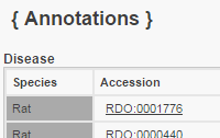
RGD Manual Disease Annotations    Click to see Annotation Detail View
TermQualifierEvidenceWithReferenceNotesSourceOriginal Reference(s)
Experimental Diabetes Mellitus  ISOEp300 (Rattus norvegicus)2312263; 2312263 RGD 
Experimental Diabetes Mellitus  IMP 2312263 RGD 
Experimental Diabetes Mellitus  ISOMef2a (Rattus norvegicus)2312263; 2312263mRNA:increased expression:heartRGD 
Experimental Diabetes Mellitus  ISOMef2c (Rattus norvegicus)2312263; 2312263mRNA:increased expression:heartRGD 
Experimental Diabetes Mellitus  IEP 2312263; 2312263mRNA:increased expression:heartRGD 

Gene Ontology Annotations    Click to see Annotation Detail View

Biological Process
TermQualifierEvidenceWithReferenceNotesSourceOriginal Reference(s)
cellular response to glucose stimulus  IEP 2312263; 2312263 RGD 
response to glucose  IMP 2312263 RGD 

Objects Annotated

Genes (Rattus norvegicus)
Ep300  (E1A binding protein p300)
Mef2a  (myocyte enhancer factor 2a)
Mef2c  (myocyte enhancer factor 2C)

Genes (Mus musculus)
Ep300  (E1A binding protein p300)
Mef2a  (myocyte enhancer factor 2A)
Mef2c  (myocyte enhancer factor 2C)

Genes (Homo sapiens)
EP300  (E1A binding protein p300)
MEF2A  (myocyte enhancer factor 2A)
MEF2C  (myocyte enhancer factor 2C)


Additional Information